Retreatment in Endodontics: What Happens When Your Root Canal Is Redone Under a Microscope
By Root Canal Foundation, Chennai
A root canal is meant to be a permanent solution. But sometimes, months or even years later, the same tooth starts hurting again, or an X-ray taken for another reason reveals a lingering infection nobody noticed. This is called a failed root canal treatment (RCT), and the good news is that it can very often be fixed — through a specialised procedure called endodontic retreatment.
At Root Canal Foundation, retreatment is one of the areas we see most often, precisely because we are known for handling cases that other clinics have found difficult to resolve. This guide walks you through what retreatment actually involves, why a microscope changes the outcome, and answers the questions patients ask us most.
Why Do Root Canals Fail in the First Place?
A root canal can fail for several reasons: a canal inside the tooth that was too narrow, curved, or hidden to be treated the first time round; a crack in the tooth; a crown or filling that let bacteria leak back in; or an infection that had already spread beyond the root tip before treatment began. Whatever the cause, the fix is rarely to simply "redo" the same procedure — it requires figuring out precisely what went wrong and correcting it, which is why retreatment is considered a more advanced skill than a first-time root canal.
This is where the dental operating microscope changes everything.
Tracing Missed Canals
Teeth do not read textbooks. Many teeth — particularly molars — have extra canals that are extremely narrow, curved, or calcified, and easy to miss without magnification. A missed canal left untreated continues to harbour infection no matter how well the other canals were cleaned, and it is one of the single most common reasons a root canal fails.
Under the microscope, magnified up to 20 times with strong, focused illumination, these hidden canals become visible: subtle colour changes in the floor of the tooth, fine grooves leading to extra openings, and calcified canals that would otherwise be invisible to the naked eye. Locating and treating these canals is often the single most important step in a successful retreatment.
Removing Instruments Left Behind
Occasionally, a small fragment of a root canal instrument separates and remains lodged inside the canal during a previous treatment. This can block proper cleaning and sealing of the canal and is a well-documented cause of persistent infection.
Removing a separated instrument is a delicate task — done without magnification, it is easy to weaken or perforate the tooth in the process. Under the microscope, using fine ultrasonic tips, the fragment can usually be loosened and retrieved with the surrounding tooth structure carefully preserved. Where a fragment cannot be safely removed, it can often still be bypassed or sealed in, so that the infection is controlled and the tooth is saved.
Apical Surgery Using Piezo Units & the Microscope
Not every failed root canal can be resolved by reopening and recleaning it from above (non-surgical retreatment). Sometimes the infection persists at the very tip of the root, or the tooth has a crown, post, or restoration that would need to be destroyed to attempt non-surgical retreatment. In these cases, a minor surgical procedure called an apicoectomy (apical surgery) is recommended.
This involves gently accessing the root tip through the gum, removing the infected tissue, and sealing the end of the root from below. Two things have transformed how predictable this surgery is:
- The microscope: Allows the surgeon to identify hairline fractures, extra canals at the root tip, and confirm a complete, clean seal under high magnification.
- Piezoelectric (piezo) ultrasonic units: Cut bone with fine, precise vibrations rather than a rotating drill. This allows the surgeon to shape a small bony window conservatively, protect nearby nerves and sinus structures, control bleeding better, and accelerate recovery.
Together, microscope-guided technique and piezo instrumentation have raised the success of apical surgery from roughly the 60% range to well above 90% in modern microsurgical practice.
Treating Large Lesions — Surgically & Non-Surgically
A long-standing infection at the root tip can grow into a cyst or lesion that shows up as a dark, well-defined area on an X-ray or 3D (CBCT) scan. Many patients assume a large lesion automatically means surgery or extraction — but that is not always the case.
- Non-surgical management: Thoroughly cleaning, disinfecting, and sealing the canal system — sometimes with the aid of medicated dressings between visits — allows even sizeable lesions to heal gradually over months as the body resorbs the infected tissue. This is always attempted first when the anatomy allows.
- Surgical management: When the lesion persists despite proper non-surgical treatment, or the canal cannot be adequately accessed from above, microscope-guided apical surgery directly removes the diseased tissue and seals the root end.
3D imaging (CBCT) plays an important role here, giving a true three-dimensional picture of the size and extent of the lesion before deciding on the right approach — something a standard 2D X-ray cannot always show accurately.
Why Experience Matters in Retreatment
Retreatment is not a routine procedure — it is one of the most technically demanding areas of endodontics, and outcomes depend heavily on the clinician’s experience and equipment.
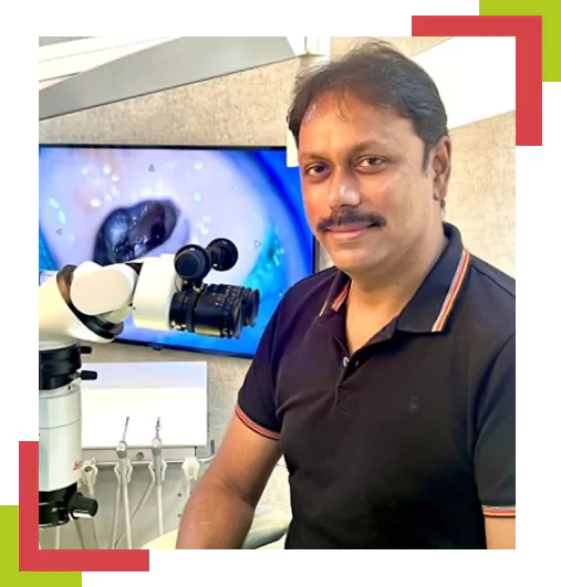
Dr. V. Gopi Krishna
MDS, FDS RCS (Edin)Dr. V. Gopi Krishna, founder of Root Canal Foundation, brings 24 years of specialist clinical experience in endodontics, with more than 25,000 endodontic cases performed, including a substantial share of complex retreatments and microsurgeries referred to him by other dentists.
He was among the first in India to bring the dental operating microscope into everyday clinical use, installing the country’s first clinical microscope in 2007. He is the author-editor of leading endodontic textbooks, including Grossman’s Endodontic Practice, read by more than 30,000 dental students across Asia each year, and holds the FDS RCS ad hominem — the highest honorary fellowship of the Royal College of Surgeons, Edinburgh — awarded to very few endodontists worldwide.
Root Canal Foundation, established in Chennai in 2004, has grown into one of India’s most reputed root canal centres, equipped with ten in-house dental operating microscopes, in-house CBCT 3D imaging, and a track record of treating referred and second-opinion cases from across India and more than 30 countries.